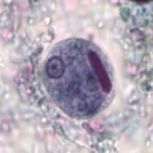

A 21-year-old exchange student from Nigeria presented to the University Clinic with a 3-day history of diarrhea, mild abdominal pain, loss of appetite, and fatigue. Stool samples collected in 10% formalin and Zn-PVA fecal preservatives were sent to the laboratory for analysis. Figures A–D show organisms seen by the technologist in a trichrome-stained slide of the Zn-PVA sample. The organisms ranged in size from 12-26 μm. What is your diagnosis? Based on what criteria? What other testing, if any, would you recommend?
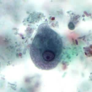
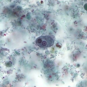
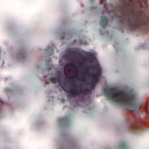
This was a case of amebiasis caused by Entamoeba histolytica/Entamoeba dispar. Diagnostic morphologic features included:
Entamoeba histolytica is morphologically identical to Entamoeba dispar and previously could only be distinguished by finding ingested erythrocytes in the trophozoites. There are reports that Entamoeba dispar is also capable of ingesting RBC during colonization, on rare occasions. As such both Entamoeba histolytica as well as Entamoeba histolytica/Entamoeba dispar are acceptable responses. It is recommended that the identification be confirmed by PCR using the Zn-PVA sample.
For more information on amebiasis please click here: https://www.cdc.gov/dpdx/amebiasis/index.html
Images presented in the dpdx case studies are from specimens submitted for diagnosis or archiving. On rare occasions, clinical histories given may be partly fictitious.